


Ectropion Correction
Advanced Surgical Solutions for Lower Eyelid Malposition
Ectropion Correction: Precise Surgical Restoration
Suffering from tearing or chronic ocular irritation due to Ectropion? Discover our precise, non-excisional surgical approach for lower eyelid reconstruction, focusing on anatomical restoration and long-term functional stability.
Understanding Ectropion
The Anatomical Shift
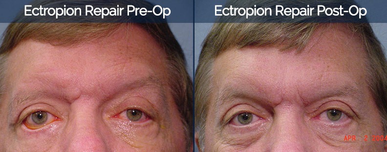
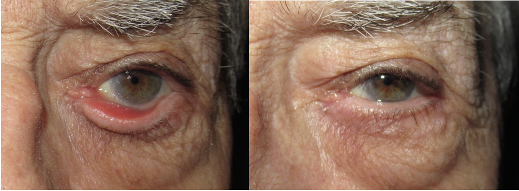
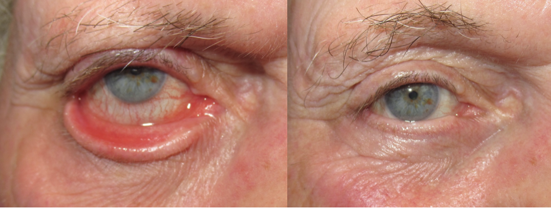
Ectropion occurs when the lower eyelid margin loses its tension and tilts outward, away from the eye. This loss of structural "angle" means the eyelid is no longer anchored correctly, leading to the exposure of the inner conjunctiva.
Why Tearing Occurs
The lacrimal punctum (tear drainage opening) relies on contact with the eye to function. Once the lid tilts outward by even 0.5 mm, the drainage system fails, causing constant tearing (epiphora) and irritation.
The Cycle of Inflammation
Persistent tearing on the skin can trigger inflammation and eczema. This leads to tissue shrinkage, which unfortunately pulls the eyelid further outward, turning a simple case into a complex deformity.
Direct Clinical Oversight
Every procedure is personally performed or directly supervised by Dr. A. Galal to ensure maximum safety.
Anatomical Precision
We focus on restoring natural eyelid function without unnecessary tissue removal or scarring.
Tailored Surgical Plans
Every patient receives a customized surgical approach based on their unique ocular anatomy and needs.
Specialized Care
Experience a refreshed, clear gaze through our dedicated, sub-specialized Oculoplastic expertise.
The Path to Oculoplastic Excellence: Our Surgical Roadmap
A Step-by-Step Approach to Safe and Precise Oculoplastic Care
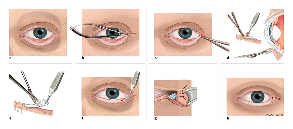
Anatomical Access
A precise approach is made to the inner surface of the eyelid to access the deeper connective tissue layers (retractors) that support the globe.
Step 01
Retractor Identification
The specialized retractor tendon sheet, which holds the structural integrity of the eyelid, is carefully isolated and prepared for re-anchoring.
Step 02
Functional Re-coupling
Using a non-excisional method, the retractor layer is precisely re-coupled with the eyelid structure, restoring the natural tension and horizontal support.
03
Step 03
Anatomical Restoration
The eyelid margin is guided back into its correct position against the globe, effectively restoring the tear drainage angle and eliminating outward rotation.
03
Step 04
BEFORE & AFTER

