
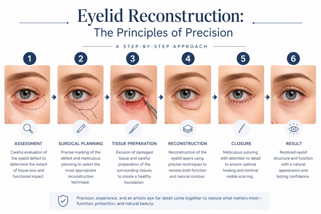
Eyelid Reconstruction
Restoring form, function, and symmetry with meticulous surgical mastery following trauma, tumor excision, or previous interventions.
The Four Concentric Zones of Complexity
Systematic surgical planning based on orbital and globe geometry.
Outside Orbital Rim
The lowest complexity zone, located outside the orbital margin, allowing for straightforward reconstruction.
Rim to Bulbus
Situated between the orbital margin and the globe, requiring precise, single-lamella management.
Equator to Limbus
The scleral zone over the globe, necessitating a robust, double-layer reconstruction approach.
Pre-Corneal Zone
Covering the cornea (such as the central third of the upper lid), representing the highest level of reconstructive complexity.
Single vs. Double-Layer Reconstruction
Anatomical precision determines structural requirements.
Zones II and III overlie the delicate surface of the globe and strictly require a robust, two-layered reconstruction to ensure both protection and mobility.
In this approach, one layer functions as a structural carrier lamella while the second layer is grafted freely. Conversely, Zones 0 and I typically require single-lamella management due to their extra-orbital positioning.
Zones 0 & I
Extra-orbital positioning allowing for single-lamella reconstruction.
Zones II & III
Pre-corneal & scleral positioning requiring a mandatory double-layer approach.
Oncologic Safety
Functional Restoration
Anatomic Precision
Expert Diagnosis
Advanced Grafting
Dedicated Aftercare
Surgical Techniques: Transposition & Transplantation
Precision tissue management tailored to anatomical geometry and surface contours.
Transposition
A tissue flap is shifted or pivoted into position while maintaining partial vascular supply through its base. While effective, this technique requires longer incision lines to reposition tissue accurately.
Free Transplantation
Tissue is completely separated from its original blood supply and grafted. Due to the exceptional vascular network of the eyelids, thin skin or split-skin grafts over a muscular bed heal with remarkable success and minimal scarring.
Concave & Convex Contour
Reconstruction accounts for spatial variations—nasal areas require tension-free concave surfaces with adequate tissue support, while temporal regions manage convex surfaces with structural precision.
Reconstruction Cases & Indications
Precision solutions categorized by origin and anatomical challenge.
Post-Tumor Excision
Restoring structural integrity and margins after oncologic resections, balancing safety with aesthetic harmony.
Post-Traumatic Defects
Realigning complex multi-layered tissue damage caused by trauma to ensure protective blink mechanics.
Post-Aesthetic Complications
Specialized revision procedures correcting functional or structural malpositions from previous cosmetic surgeries.
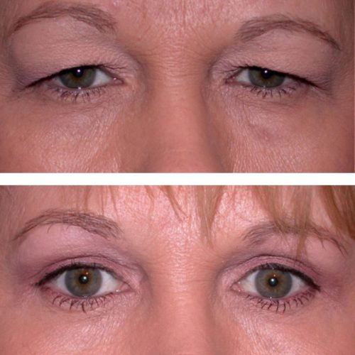
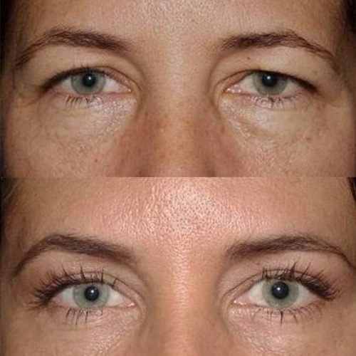
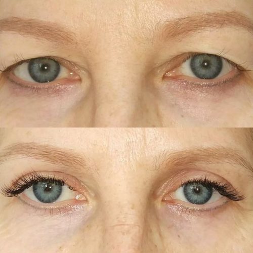
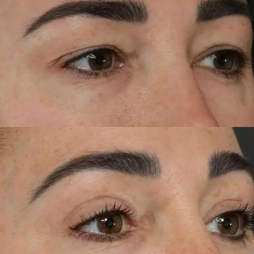
BEFORE & AFTER