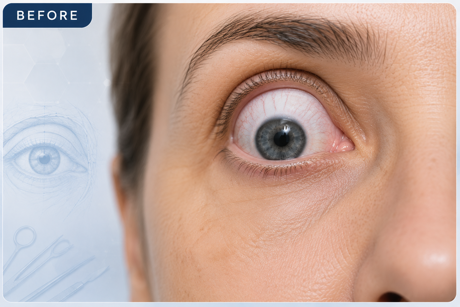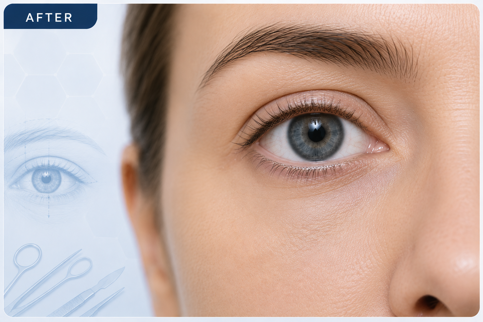

Eye socket (orbita)
Exploring the complex relationship between eyelid and orbital surgery, highlighting minimally invasive transpalpebral and transconjunctival techniques for seamless access to all orbital quadrants and structural repair.
Orbital Surgery & Integrated Oculoplastic Approaches
The specialty of oculoplastic surgery is intrinsically linked to orbital surgery. By utilizing transpalpebral and transconjunctival entry points, surgeons can access all orbital quadrants through postoperatively invisible incisions while preserving complex fibroelastic structures.
Fibroelastic System & Orbital Fat Compartments
Understanding the elastic structures and fat pad distributions that define surgical boundaries during orbitotomy and blepharoplasty.
Fibroelastic Support Architecture
Originating from Tenon's Capsule (Fig. 1)The elastic framework of the orbit originates from Tenon's capsule (cingulate ligament), providing suspension, structural integrity, and guided movement for ocular structures.
-
Tenon's Capsule: The main fibrous envelope anchoring muscle insertions and orbital fascia.
-
Tendon Arches: Crucial supportive ligaments including Whitnall's ligament (superior) and Lockwood's ligament (inferior).
-
Radial Septa: Fibrous walls dividing the movable orbital fat into distinct anatomical compartments.
Orbital Fat & Glandular Anatomy
Movable Fat Pads & Tissue Boundaries (Fig. 2)Movable orbital fat lies suspended between the radial septa, protecting the globe and acting as key surgical landmarks:
Orbitotomy Access & Quadrant Strategies
Surgical entry routes utilizing natural eyelid folds and transconjunctival planes for complete orbital exposure with minimal aesthetic footprint.
Medial Orbitotomy
Access to Medial Quadrants & Orbital Floor
Lateral & Inferior Orbitotomy
Access to Lateral Quadrants & Central Floor
Complete Orbital Coverage (360° Reach)
By combining both the medial upper crease approach and the transconjunctival lower lateral approach, surgeons can safely reach all quadrants of the orbit (Figs. 3–6).
BEFORE & AFTER