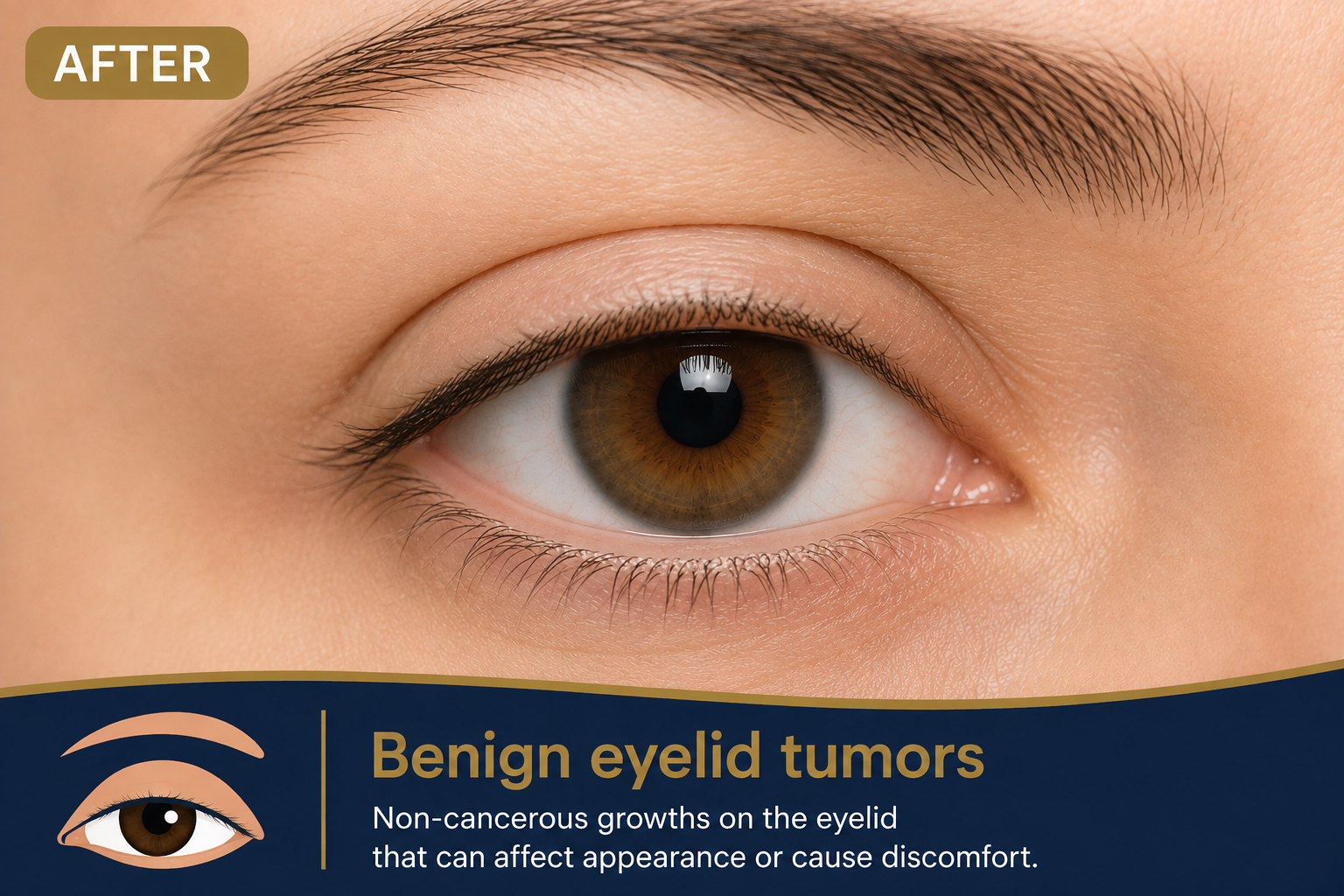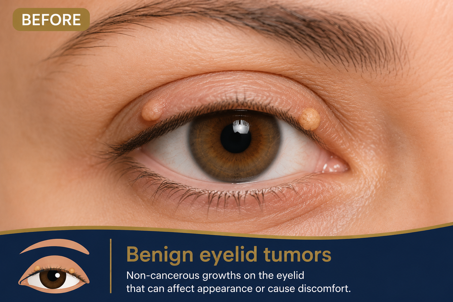

Benign eyelid tumors
Clinical insights into diagnosing and differentiating benign eyelid tumors and cysts using biomicroscopy, featuring common lesions like Xanthelasma, Molluscum, and Keratoacanthoma.
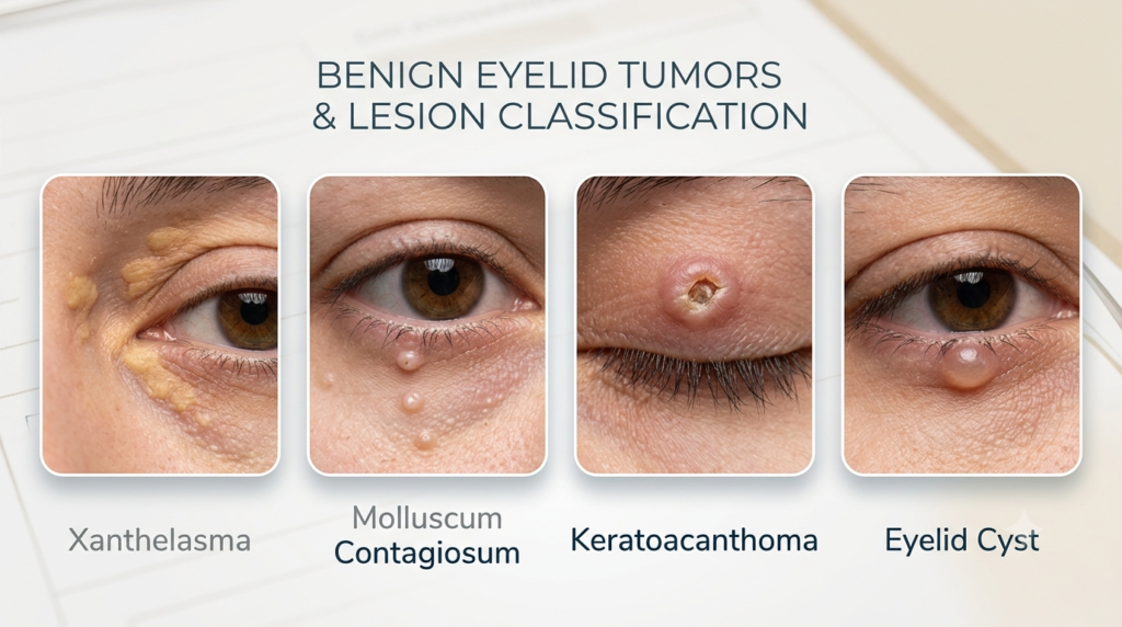
Benign Eyelid Tumors & Lesion Classification
Accurate differentiation from malignant processes is paramount. 80%–90% of eyelid tumors can be classified using slit lamp biomicroscopy. This involves evaluating cysts, pigmented lesions (nevi, basal cell carcinomas), and diverse granulomatous contents.
Common Examples of Benign Eyelid Lesions
Cyst formation is almost always a benign process. Cysts can accumulate a variety of liquid, solid, or granulomatous contents.
Xanthelasma
Sharply demarcated yellowish deposits of cholesterol underneath the skin. While benign, they may indicate underlying lipid disorders.
Molluscum Contagiosum
Small, pearly, dome-shaped papules with a central umbilication. Viral etiology, common in children or immunocompromised adults.
Keratoacanthoma
Rapidly growing, solitary epithelial tumor, often with a keratin-filled crater. Histologically benign but can mimic squamous cell carcinoma.
Malignant Potential & Differentiation Challenges
Tumors initially assessed as benign can, in rare cases, become malignant. Differentiation can be difficult in some cases, highlighting the critical importance of specialist evaluation and potential biopsy.
BEFORE & AFTER