

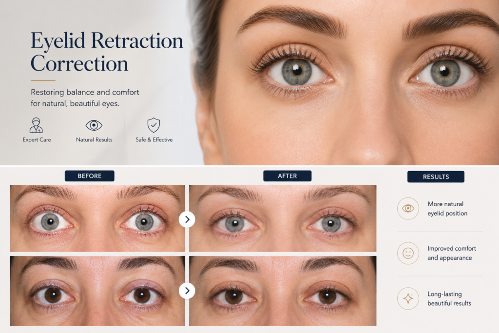
Eyelid retraction
Abnormal elevation or depression of the eyelid caused by tissue loss, scarring, or underlying conditions like Graves’ disease.
Pre-Surgical Evaluation & Diagnostics
Essential diagnostic priorities required before planning surgical correction for eyelid retraction.
Lamella Identification
Determine which layer (anterior, posterior, or both) causes retraction.
Cicatricial Traction
Assess scar pull and postinflammatory tissue fibrosis.
Surgical History
Evaluate previous eyelid surgeries or trauma-induced defects.
Motility Check
Verify whether independent movement of lid layers is inhibited.
Spacer Function
Plan the use of orbital fat to prevent transversal scar adhesions.
Etiology Screening
Rule out systemic factors such as Graves' (Basedow's) disease.
Types of Lid Retraction
Understanding whether a retraction is simple or complex is critical before planning any surgical correction.
Single Layer Involvement
In simple retraction cases, either the anterior or the posterior lamella is affected independently, allowing for a more straightforward surgical approach.
Multi-Layer Complicated Retraction
Often resulting from trauma, penetrating injuries, or conditions like Basedow's disease (endocrine retraction), involving multiple layers and inhibiting independent movement.
Key Clinical Presentations
- Complicated Lower Lid Retraction: Following orbital floor trauma involving all eyelid layers.
- Complicated Upper Lid Retraction: Post-traumatic presentation with multi-layer structural involvement.
- Endocrine Lower Lid Retraction: Associated with Graves' / Basedow's disease pathologie.
BEFORE & AFTER
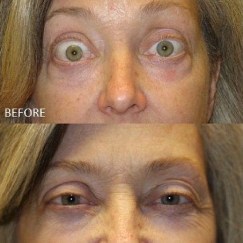
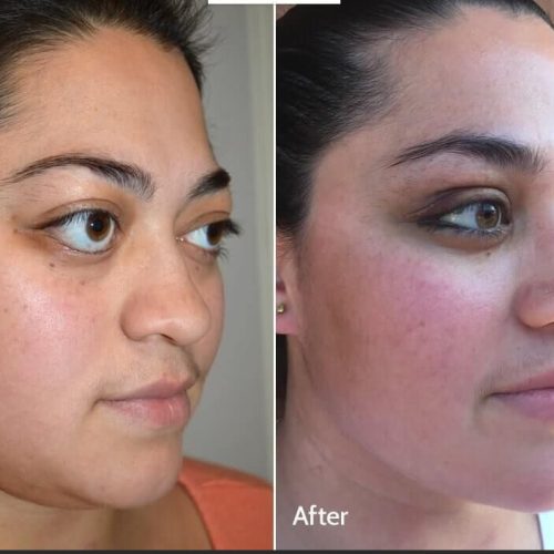
Examples of Simple or Complicated Lid Retraction
A clinical overview demonstrating how different pathologies result in specific patterns of eyelid tissue retraction.
Post-Traumatic Lower Lid Retraction
Involves complete structural scarring where ALL eyelid layers (anterior and posterior) are affected, inhibiting independent lamellar movement.
Post-Traumatic Upper Lid Retraction
Complex multi-layer involvement demanding advanced reconstructive techniques, often utilizing orbital fat as a gliding spacer.
Graves' Disease Lower Lid Retraction
While potentially complex, it often presents with a more localized, single-layer simple retraction pattern amenable to targeted surgical correction.
The Gliding Layer Mechanism
How orbital fat serves as a dynamic tissue spacer to protect postoperative lid movement.
Preserving Independent Lid Movement
In complex retractions, scarring binds the anterior and posterior lamellae together, locking eyelid motion. Re-establishing the anatomical gliding plane is crucial.
Transversal Scar Prevention
Orbital fat is strategically positioned between reconstructed layers to block restrictive fibrotic adhesion formation.
Anatomical Spacer Function
Acts as a natural buffer, ensuring the front and back lamellae slide smoothly and independently during blinking.