
Ptosis
Abnormal drooping of the upper eyelid caused by levator muscle weakness, nerve impairment, or congenital underdevelopment.
Ptosis (Upper Eyelid Drooping)
Understanding true ptosis requires a specialized anatomical approach. It is not merely loose skin, but an internal structural weakness of the eyelid levator muscle.
What is True Ptosis?
Ptosis occurs when the upper eyelid sits abnormally low on the eyeball, covering the pupil partially or completely. True ptosis operates independently of outer skin changes and cannot be corrected with a standard external "eyelid lift" (blepharoplasty)—it requires precise surgical treatment of the levator palpebrae superioris muscle within the eye socket.
Pre-Surgical Diagnostic Priorities
Ptosis can stem from nearly 100 different causes. Identifying the precise underlying mechanism is essential before planning surgical levator repair.
Origin Classification
Distinguishing congenital cases (associated with levator dystrophy) from acquired forms.
MRD Assessment
Measuring the Marginal Reflex Distance to categorize severity from mild to negative MRD.
Levator Function (LF)
Evaluating vertical lid excursion (normal 15–16 mm) across elevation and depression.
Bell's Phenomenon
Testing upward globe rotation during closure to ensure adequate postoperative corneal protection.
Orbicularis Function
Verifying the strength of eyelid closure and blinking dynamics to prevent exposure risk.
Spontaneous Stimulation
Checking central visual system drive, ruling out amblyopia or neural pathways issues.
Classification of Ptosis
All cases of ptosis are broadly categorized based on their onset (Congenital vs. Acquired) and underlying complexity.
Acquired & Simple Ptosis
Non-congenital form affecting only the anatomy of the eyelid levator muscles.
- Structural weakness or detachment of the levator aponeurosis.
- Preserved neuromuscular closure mechanisms and normal motility.
- Highly predictable outcomes with targeted levator reattachment surgery.
Congenital Ptosis
Present from birth due to developmental anomalies of the lifting muscle.
- Characterized by dystrophy or underdevelopment of the levator palpebrae superioris.
- Limited muscle function causing both elevation and depression deficits.
- May present with typical signs like lid lag and lipodystrophy.
Complicated Ptosis
Rare acquired or congenital forms with secondary neuromuscular involvement.
- Defect rooted deep in the muscular or nervous system (neurogenic/myogenic).
- Impaired eye closure mechanisms and associated ocular motility disorders.
- Requires expanded diagnostic workup and customized complex repair.
Clinical Measurement & Diagnostic Protocols
Precise diagnostic measurements are mandatory to dictate the exact surgical technique for lifting the eyelid.
Marginal Reflex Distance (MRD)
Corneal reflection to upper eyelid marginNormal MRD ranges between 2.5 mm and 4.5 mm. Severity is graded based on deviation from normal:
Levator Function & Ocular Mechanics
Evaluating muscle excursion and ocular protectionLevator Function (LF = Elevation + Depression)
Vertical lid movement between maximum downward and upward gaze. Normal function is 15–16 mm. In congenital cases, both elevation and depression deficits occur, whereas simple acquired ptosis preserves depression dynamics.
Key Corneal Protection Tests
- Bell's Phenomenon: Spontaneous upward rotation during closure. If absent, expanded complex diagnostics are required.
- Orbicularis Oculi Test: Verifying closure strength to prevent post-op exposure keratitis.
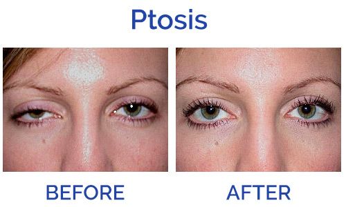
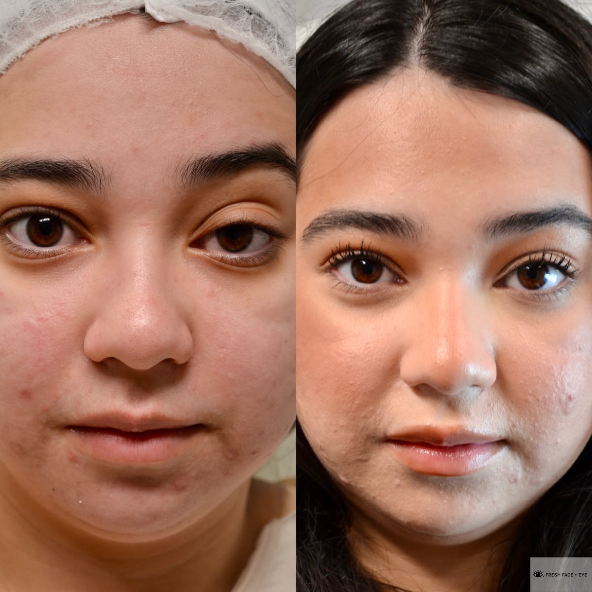
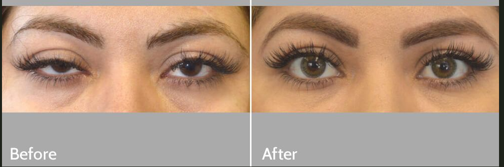
BEFORE & AFTER