
Malignant eyelid tumors
Clinical insights into diagnosing and differentiating benign eyelid tumors and cysts using biomicroscopy, featuring common lesions like Xanthelasma, Molluscum, and Keratoacanthoma.
Malignant Eyelid Tumors: Diagnosis & Management
Most malignant eyelid tumors can be identified and classified clinically via biomicroscopy (slit lamp) without immediate tissue sampling. Mastering advanced surgical techniques is crucial to avoid incomplete removal, ensuring sufficient safety margins while employing diverse reconstruction strategies.
Mohs Micrographic Surgery & Margin Control
The Mohs technique offers crucial control over tumor-free resection margins, utilizing systematic histological examination of tissue strips.
Systematic Resection Protocol
0.5 mm Strips & 3 mm Safety MarginFollowing the main specimen resection (0) with a 3 mm safety margin outside visible borders, narrow 0.5 mm margin strips (1,2,3,4) are obtained for histology.
-
Marginal Excisions: Mohs technique excisions performed at four distinct edges of the tumor.
-
Orientation Marking: Strips are harvested, marked, and examined individually for residual tumor.
-
Subcutaneous Awareness: Critical for recurrences, which tend to grow subcutaneously and may be present even when medially invisible (Fig. 3–5).
Tumor-Specific Safety Margins
Resection standards by pathologyThe safety margin outside macroscopic borders varies based on histological classification to ensure complete eradication:
Eyelid Reconstruction & Defect Management
Advanced restorative techniques tailored to defect size following complete tumor removal and margin verification.
Pentagonal Resection
Pentagonal resection and direct transposition of the eyelid remnant is highly effective when the surgical defect accounts for approximately one-third of the total eyelid area.
Lateral Cantholysis
For somewhat larger defects, the lateral cantholytic ligament can be divided (cantholysis). This procedure successfully loosens the remaining lateral eyelid to achieve primary closure.
Right-Angled Resection
In cases of larger structural defects, a right-angled resection is performed, allowing the posterior and anterior lamellae of the eyelid to be reconstructed separately for optimal functional restoration.
Surgical Mastery & Preservation
Mastering these varied reconstruction methods ensures that surgeons achieve oncologically safe margins without compromising eyelid functionality or tissue alignment.
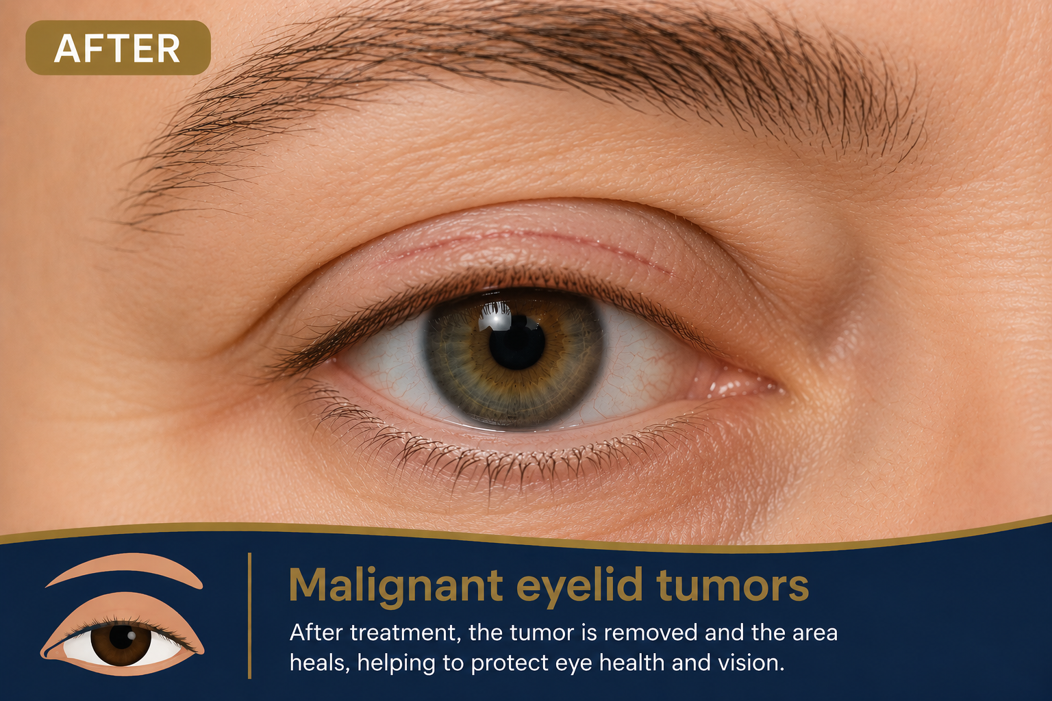
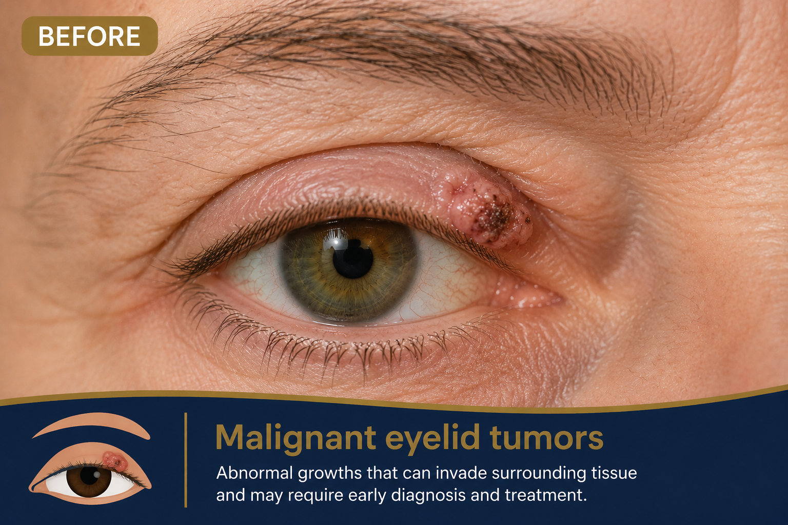
BEFORE & AFTER